Scientists have created a detailed atlas of the human female reproductive tract during fetal development, thanks to a newly published study led by Dr. He and her colleagues.
Mapping fetal development
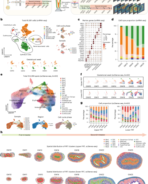
The researchers used a combination of microscopy, computational modeling, and machine learning to create a 3D visualization of the developing reproductive tract from 10 to 25 weeks of gestation.
The team analyzed thousands of tissue samples from human fetuses, applying advanced algorithms to identify and classify the distinct segments of the reproductive tract.
Breaking down the process
The study found that the female reproductive tract is composed of multiple segments, each with its own unique transcriptome – the complete set of RNA transcripts produced by the genome.
The researchers used single-cell RNA sequencing to identify and characterize the specific cells present in each segment, laying bare the intricate details of fetal development.
According to the study, the segments of the female reproductive tract undergo significant changes in development, with some regions exhibiting rapid growth and others showing slower development.
What this means
The research has significant implications for our understanding of reproductive health, particularly when it comes to the development of medical treatments and interventions.
By creating a detailed atlas of fetal development, researchers can better identify potential issues and develop targeted therapies to improve reproductive outcomes.
The study’s findings may also have applications in the field of regenerative medicine, where researchers aim to develop therapies that can repair or replace damaged tissues.